Yolanda Carrascal, Jaime Arroyo, Horacio Valenzuela y Mireia Fernandez
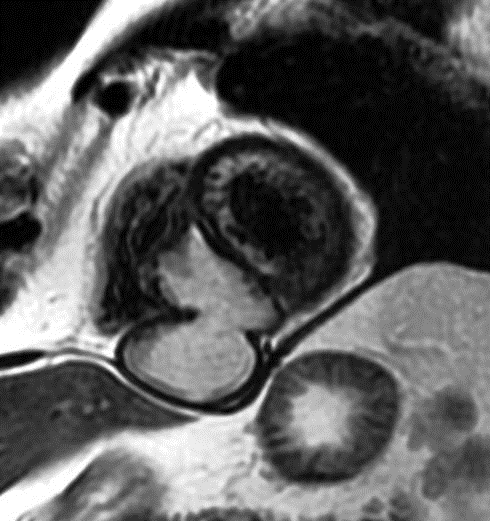
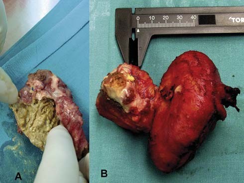
A 60-year-old male presented with pulmonary hydatid cyst excision (after vomica with hemoptysis) when aged 20 years. He was referred with a cystic mass in interventricular septum, in magnetic resonance imaging (MRI) (Fig. 1). Due to high suspicion of hydatic cyst (it can remain latent up to five decades), surgical treatment was decided (Fig. 2).
Figure 1: After a syncopal episode, MRI identified a bilobulated cystic mass in interventricular septum, externally protruding to ventricular diaphragmatic surface, and extending upwards membranous septum (48.54 × 65.63 mm).Figure 2: (A) Open hydatic cyst with ‘caseum’ content. Neither daughter cyst nor scoleces or cuticular membrane was identified. Pathological analysis described cyst content as a ‘caseum’, probably secondary to degenerative hydatic cyst necrosis. (B) Complete hydatic cyst after surgical resection.